Easter Sunday

Easter Sunday

About Me

My photo
Newburgh, Indiana, United States

Monday, April 7, 2008

Glomus Caroticum Tumor

The most common type of paraganglioma is the glomus caroticum tumor within the neck. They are slow growing tumors but can double in size within 4 years. Six to twelve percent of the cases are malignant. Given their location they are not the easiest to get to and are risky to perform surgery on. There are 3 treatment options that include surgery, radiation therapy, and just watch and see if it gets bigger. The cure rate for these tumors ranges from 89 to 100 percent however, 20 percent of them have cranial nerve damage. If the tumor grows to be 5cm or more intervention is considered. Risks of surgery include bleeding out and stroke from clamping the carotid artery. For very large tumors, multiple tumors and patients that are not good candidates for surgery, or due to the patients age, may require radiation therapy rather than surgery.

Reference:
http://www.ispub.com/ostia/index.php?xmlFilePath=journals/ijtcvs/vol10n2/nodule.xml

Tuesday, April 1, 2008

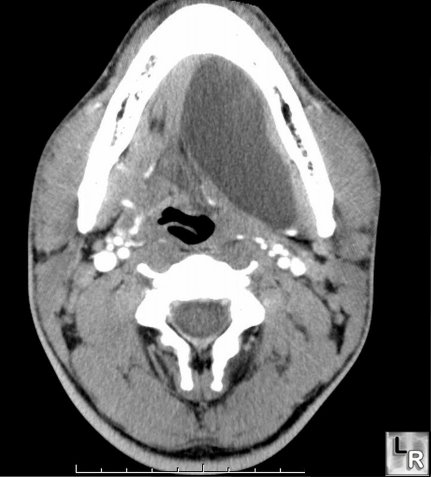
Ranula

Ranulas are termed as pseudocyts that are caused by blockage of flow of saliva or can be caused by trauma. Ranulas usually arise from the major salivary glands that can be found in the oral cavity or in the cervical area. Ranulas account for .2% per 1000 people. According to the Minnesota Oral Disease Prevalence Study randula occurrences are ranked 41st. They account for 6% of oral sialocysts. Ranulas on the cervical are considered rare. Ranulas are usually painless and are typically not life threatening. However, they may interfere with swallowing, chewing, speech and respiration functions. They tend to be more prevalent in males. Oral ranulas are usually under the tongue and are a bluish tint and can medially and superiorly displace the tongue. Causes can include traumas from birth from forceps or suction of the infants mouth, sucking on fingers, passing through the birth canal. Escaped mucus from an injured gland causing the raised bump. Treatment includes lancing the ranula and draining the fluid, surgical excision including the salivary gland associated, or an intracystic injection.


Refrences:

www.rad-pb.de/e1543/e1546/e2109/e2110/ranula.jpg

http://www.emedicine.com/derm/topic648.htm

http://www.learningradiology.com/caseofweek/caseoftheweekpix2/cow146.jpg

Sunday, March 23, 2008

Moyamoya Disease


Moyamoya disease is a condition where there is progressive blockage or occlusion of the ends of the internal carotid arteries and their major "terminal" branches in the brain. On a cerebral angiogram a classic sign is a puff of smoke appearance which is actually a bunch of tiny vessels due to the blockage. The term moya means tiny in Japanese. This disease affects children as well as adults. The main cause of the disease is unknown but there are some other diseases associated with moyamoya including some neurocutaneous syndromes (like meningitis, down syndrome and others). Symptoms in children are strokes, and seizures. In adults symptoms include brain hemorrhage and strokes brought on by headaches neurological impairments, and sudden collapse. If the hemorrhage is deep within the brain it is termed an intraparenchymal hemorrhage. To diagnose the disease a CT scan, a MRI, or a MRA to view the vessels in the brain. For treatment of this disease surgery is the preferred method. The surgery may include removal of the hematoma in the brain, clipping an aneurysm, or doing a brain bypass.
Reference:

Tuesday, March 18, 2008

Angiofibroma

Angiofibroma, often referred to as juvenile angiofibroma, is a beguine tumor within the nasal cavity. It is mostly common in males up to the age of 15. It can grow to be so large that it may distort and displace surrounding tissues. CT or MRI can display the location of the tumor and angiography is used to visualize any vessels that are supplying blood to it, which usually is the maxillary artery. Because of the tumors high vascularity it is usually not biopsied. Surgically removing the tumor is preferred and preoperative embolisation has been shown to significantly reduce blood loss during the procedure. Radiotherapy can be used for tumors that are risky, hard to reach, or recurrent. Hormonal treatment may also become a possibility. Symptoms include nosebleeds, pressure, mass appearing under the skin on the cheek, and may cause vision problems. The culprit of the condition is unknown but suspected to be because of the release of sex hormones.


References:


Tuesday, February 26, 2008

Graves Disease

Graves disease is an autoimmune caused from an overactive thyroid (hyperthyroidism). This disease tends to effect women within the age of 20 to 40, however it is found in children, the elderly and infants. Symptoms include heat sensitivity, goiters, bulging of eyes, fatigue, sleeplessness, weight loss, and irritability. Patients may also develop red thickening of the skin on the shins called pretibial myxedema. Patients may not develop symptoms at all. Some things that have been linked for the cause of this disease is possible heredity, stress, hormones levels, age, sex and body's immune system. There are three treatments for this disease including radioactive iodine, medication, and surgery. If left untreated it may cause problems with pregnancy and may even lead to death.

Tuesday, February 19, 2008

Macroadenoma

Pituitary Macroadenoma is a non-cancerous tumor on the pituitary gland that has grown more than 10mm. Since the pituitary gland produces a number of hormones that regulate differnt things there are different classifications of macroadenoma depending on the symptoms and hormones sectreted. If the pituitary stops producing hormones it is termed a non-functioning pituitary macroadenoma. Some symptoms are specific to certian macroadenomas.

Symptoms range from mild, tiredness or restlessness, to severe like headaches, vomiting and/or dizziness. If the macroadenoma becomes large enough it can press on the optic nerve causing vision complications. Large pituitary macroadenomas can distort the sella turcica making it larger than normal. They can also cause midline shifts and depress on different portions of the brain causing varying symptoms.

(Image showing enlarged sella turcica)

Diagnosis is usually obtained by asking the patient for any family history of the disease, symptoms the patient is having, and if the patient is on any medication. Both CT and MRI exams can be ordered to view the pituitary gland.


Treatment depends on the size, shape and type of the tumor as well as symptoms and overall health of the patient. Treatment ranges from surgery, radiation therapy and drg therapy.


References:

http://endocrine-system.emedtv.com/pituitary-macroadenoma/pituitary-macroadenoma-p3.html


www.pathology.vcu.edu/.../images/1-2.jpg

Sunday, February 10, 2008

Acoustic Neuroma


Acoustic Neuroma is a non-cancerous tumor that is found in the IAC from the vestibulo-cochlear nerve. Since this nerve is responsible for balance and hearing symptoms include hearing loss, ringing in the ears, vertigo, difficulty balancing, pressure in the ear and with larger tumors numbness in the face. Diagnosing AN is done by a hearing test called and auditory brainstem response which records responses from the brain stem. Imaging technology has made it easier to diagnose ANs. Both CT and MRI can be used but MRI is preferred with the use of gadolinium. Options for treatment range from observation to radiation therapy. They also consider microsurgical removal. Because they are normally benigne microsergery is usually the last resort to releave pressure and to improve hearing. If the tumor gets to big then symptoms may increase and worsen. ANs are mostly diagnosed in women around 30 to 60 in age and 2.5% of the population is speculated to have small non-symptomous tumors.


References: